How cells learn to ‘count’
One of the wonders of cell biology is its symmetry. Mammalian cells have one nucleus and one cell membrane, and most humans have 23 pairs of chromosomes. Trillions of mammalian cells achieve this uniformity — but some consistently break this mold to fulfill unique functions.

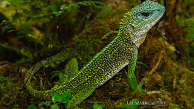
The surface of a multicillated cell. / Photo: Andrew Holland
EurekAlert | JOHNS HOPKINS MEDICINE
Listen to this article
Leer en español: Cómo las células aprenden a ‘contar’
Now, a team of Johns Hopkins Medicine researchers has found how these outliers take shape.
In experiments with genetically engineered mice, a research team has ruled out a mechanism that scientists have long believed controls the number of hairlike structures, called cilia, protruding on the outside of each mammalian cell. They concluded that control of the cilia count might rely instead on a process more commonly seen in non-mammalian species.
The experiments, described Dec. 2 in Nature Cell Biology and led by Andrew Holland, Ph.D., associate professor of molecular biology and genetics at the Johns Hopkins University School of Medicine, may eventually help scientists learn more about human diseases related to cilia function, such as respiratory infections, infertility, and hydrocephaly.
Cilia are ancient structures that first appeared on single-celled organisms as small hairlike "fingers" that act as motors to move the cell or antennae to sense the environment. Nearly all human cells have at least one cilium that senses physical or chemical cues. However, some specialized cell types in humans, such as those lining the respiratory and reproductive tracts, have hundreds of cilia on their surface that beat in waves to move fluids through the system.
"Our main question was how these multicilliated cells become so dramatically different than the rest of the cells in our body," says Holland. "Most cells make exactly one cilium per cell, but these highly specialized cells give up on this tight numerical control and make hundreds of cilia."
In an effort to answer the question, Holland and his team took a closer look at the base of cilia, the place where the organelles attach and grow from the surface of the cell. This base is a microscopic, cylinder-shaped structure called a centriole.
In single-ciliated cells, Holland says, centrioles are created before a cell divides. A cell contains two-parent centrioles that each duplicate so that both new cells gets one pair of centrioles — the oldest of these two centrioles then goes on to form the base of the cilium. However, multicilliated cells create unique structures, called deuterostomes, that act as a copy machine to enable the production of tens to hundreds of centrioles, allowing these cells to create many cilia.
"Deuterostomes are only present in multicilliated cells, and scientists have long thought they are central for determining how many centrioles and cilia are formed," says Holland.
Also read: Science's 2019 breakthrough of the year: the first image of a black hole
To test this, Holland and his team developed a mouse model that lacked the gene that creates deuterostomes. Then, they analyzed the tissues that carry multicilliated cells and counted their cilia.
The researchers were surprised to find that the genetically engineered mice had the same number of cilia on cells as the mice with deuterostomes, ruling out the central role of deuterostomes in controlling the number of cilia. For example, the multicilliated cells lining the trachea all had 200-300 cilia per cell. The researchers also found that cells without deuterostomes could make new centrioles just as quickly as cells with them.
With this surprising result in hand, the researchers engineered mouse cells that lacked both deuterostomes and parent centrioles and then counted the number of cilia formed in multicilliated cells.
"We figured that with no parent centrioles and no deuterostomes, the multicilliated cells would be unable to create the proper number of new cilia," says Holland.
Remarkably, Holland says, even the lack of parent centrioles had no effect on the final cilia number. Most cells in both normal and genetically engineered groups created between 50 and 90 cilia.
"This finding changes the dogma of what we believed to be the driving force behind centriole assembly," explains Holland. "Instead of needing a platform to grow on, centrioles can be created spontaneously."
While uncommon in mammals, the so-called de novo generation of centrioles is not new to the animal kingdom. Some species, such as the small flatworm planaria, lack parent centrioles entirely and rely on de novo centriole generation to create the cilia they use to move.
Also read: How can healthcare achieve real technology-driven transformation?
In further experiments on genetically engineered mice, Holland found that all the spontaneously created centrioles were assembled within a region of the cell-rich with fibrogranular material — the protein components necessary to build a centriole.
He says he suspects that proteins found in that little-understood area of the cell contain the essential elements necessary to construct centrioles and ultimately control the number of cilia that are formed. Everything else, the deuterostomes and even the parent centrioles are "not strictly necessary," he says.
"We think that the deuterostomes function to relieve pressure on the parent centrioles from the demands of making many new centrioles, freeing up parent centrioles to fulfill other functions," says Holland.
A better understanding of mechanisms that limit cilia number in human cells could potentially advance efforts to treat cilia-related disorders, he said, by identifying targets for drugs.